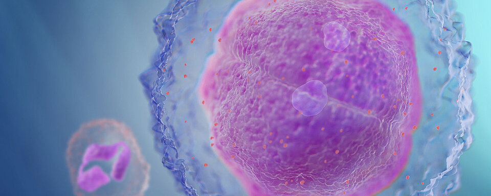
Reticulocyte haemoglobin equivalent – RET-He
What is RET-He – the Reticulocyte haemoglobin equivalent?
Measuring the haemoglobin content of reticulocytes, also known as RET-He or reticulocyte haemoglobin equivalent, is a way of diagnosing and monitoring iron deficiency anaemia. RET-He is the fastest way to detect changes in iron status. Since red blood cells have a 120-day lifetime, detecting iron deficiencies and changes in the iron status of erythropoiesis is only possible relatively late using classical haematological parameters such as HGB, MCV, MCH, or by measuring hypochromic red blood cells (HYPO-He).
Reticulocytes, the precursors of mature red blood cells, are swept into the blood stream from the bone marrow and usually mature over the course of two to four days. Measuring the number of reticulocytes is therefore a quick measure of ‘quantity’ in erythropoiesis in the marrow. Measuring the haemoglobin content of the reticulocytes means you can look at the current iron supply to erythropoiesis and judge the ‘quality’ of the cells. This lets you detect changes in iron status far earlier than through the haemoglobin content of mature red blood cells.
Why is RET-He a more effective marker?
In general, functional iron deficiency refers to the failure to release iron rapidly enough to keep pace with the demands of the bone marrow for erythropoiesis. It may occur even when the body has adequate iron stores. Measuring the haemoglobin content of the reticulocytes as a direct assessment of the iron actually used for the biosynthesis of haemoglobin can indicate whether there is enough iron available for erythropoiesis. It takes a snapshot of the ‘quality’ of erythropoiesis and is an important tool for diagnosing and monitoring iron deficiency diseases.
Conventional biochemical markers for assessing iron status, such as serum iron, transferrin or ferritin, are so drastically disturbed during inflammation with an acute phase response, or in the presence of many other severe diseases, that a clinical interpretation of the results is difficult or impossible.
For example, while low ferritin levels unequivocally indicate a lack of iron, normal or elevated levels do not let you draw any conclusions as to the bioavailability of the iron. In the presence of chronic diseases such as rheumatoid arthritis, but also in the presence of liver damage, tumours or chronic kidney disease, ferritin can also be elevated in the case of functional iron deficiency.
Benefits
The clinical utility of the Ret-He parameter has been proven and it is now an established parameter in advanced haematological analysis. ‘Reticulocyte haemoglobin content’ is recommended in nephrology guidelines such as the European Best Practice Guidelines (EBPG), National Kidney Foundation Kidney Disease Outcome Quality Initiative (NKF KDOQI).
Ret-He:
- Considered the most sensitive indicator of current iron availability for erythropoiesis
- RET-He and RET# together let clinicians draw conclusions on both the quality and quantity of the young RBC fraction.
- Is an early marker for disease - earlier than clinical chemistry markers!
- Fast, inexpensive and practical to perform!
Who/which organisation benefits from using RET-He?
Anaemia is a common symptom of several diseases and one of the most underestimated red blood cell disorders. As a result, knowing a patient’s erythropoietic status can be essential. All our analysers equipped with the RET channel offer the RET-He parameter as a diagnostic reportable.
- It is often used for patients with nephrological (kidney) disorders as they frequently suffer from anaemia in parallel. It is therefore especially important to include patients from the nephrology department or patients from dialysis centres and practices in analysis.
- Ret-He is important for patients with anaemia of chronic disease (ACD). Any patient with a chronic inflammatory process, chronic infection or malignancy can develop ACD.
- Patients with iron deficiency anaemia (IDA) will also benefit. IDA is widespread, underdiagnosed and can be found in a variety of patients. Some paediatric patients are vulnerable to developing IDA due to the growth phase.
Using RET-He
RET-He alone gives information on the current bioavailability of iron – a low value means iron is lacking or iron is not bioavailable for erythropoiesis. It is often used together with ferritin. Since ferritin is increased during the acute phase of diseases, inflammation should be ruled out, e.g. by CRP.
- a high or normal ferritin value together with a low RET-He value can suggest functional iron deficiency
- low ferritin values together with low RET-He suggest a classic iron deficiency.
- RET-He is used for monitoring erythropoietin (EPO) and/or IV iron therapy. If the value increases it indicates the therapy is having a positive effect.
RBC-He and Delta-He
While RET-He is a measure of the haemoglobin content in reticulocytes, the new diagnostic parameter RBC-He delivers information about the haemoglobin content of mature red blood cells. The parameter Delta-He is derived from the difference between these two parameters.
Delta-He values above the normal range may indicate an improvement in erythropoiesis, for example after EPO and/or iron therapy. In contrast, when Delta-He values are below the normal range for longer periods, this may indicate the onset of an anaemia.
Scientific Literature
Dr. Margreet Schoorl: The clinical use of Ret-He in the differential diagnosis of anaemia.
The clinical use of Ret-He in the differential diagnosis of anaemia.
Dr. Margreet Schoorl
Department for Clinical Chemistry, Haematology and Immunology
Northwest Clinics
Alkmaar
The Netherlands
In case of anaemia microcytic erythropoiesis is frequently due to iron deficiency and alfa- or beta-thalassaemia. Iron-deficient erythropoiesis (IDE) and thalassaemia are both associated with mild to moderate microcytic anaemia, which frequently leads to an incorrect diagnosis. It is important to discriminate between iron-deficiency anaemia (IDA) and thalassaemia, and to avoid unnecessary iron therapy to prevent development of haemosiderosis, which may result in serious complications like cardiomyopathy, liver fibrosis or endocrine dysfunctions.2,3
A wide range of laboratory parameters is available for anaemia screening and assessment of iron status. However, no single marker or combination of tests is optimal for discrimination between iron deficiency, functional iron deficiency and thalassaemia. The available indicators do not provide sufficient information and must be used in combination to obtain reliable information. In addition, iron deficiency often occurs in combination with other diseases which complicates the diagnosis. Diagnosing subjects with combined thalassaemia minor and iron deficiency is even more challenging.
Red blood cell and reticulocyte haemoglobin content
In the past years new parameters for eythropoiesis became available for investigation of the
haemoglobin content of (immature) red blood cells and for the production of red blood cells. Haemoglobin content in reticulocytes (Ret-He) is a sensitive indicator for monitoring short term deteriorations in iron and vitamin availability for erythropoiesis. Reticulocyte maturation coincides with progressive decrease in RBC volume and Hb content. If compared with the interpretation of haemoglobin content of mature RBCs (RBC-He), the interpretation of haemoglobin content of reticulocytes yields additional information concerning various states of decreased availability of iron for the haemoglobinisation of erythroid precursors.4,5 If iron supplements are administered in subjects with depleted iron stores, Ret-He already increases within a few days.6
In normal circumstances results of Ret-He are approximately 10% higher than RBC-He. Delta-He (=Ret-He minus RBC-He) results below the reference range indicate poor erythropoiesis. In contrast, results above the reference range indicate normal or improved erythropoiesis.7
Subjects with microcytic anaemia
In subjects with microcytic anaemia and suspected IDA or thalassemia, results of Ret-He and RBC-He results are both decreased if compared with a group of apparently healthy subjects.
However, using a combination of Ret-He and Delta-He a clear distinction can be made between subjects with iron deficiency and thalassemia.
In case of iron deficiency, a decreased Ret-He (cut-off value <1850 aMol) is demonstrated in combination with a decreased Delta-He. In the presence of thalassemia, usually a decreased Ret-He with a normal Delta-He is demonstrated.
In summary, the laboratory screening for anaemia has been improved by using the new haemocytometric parameters and by the development of new discriminating algorithms for the diagnosis of IDE and thalassemia.8,9
To conclude:
We strongly advise the discriminating parameters for anaemia discrimination in order to reduce diagnostic testing for confirmation and to proper diagnose the underlying cause(s) in several categories of patients, such as subjects with microcytic anaemia, women in the third trimester of pregnancy, adolescents and elderly people (age >75 year).
References
1 De Benoise B, McLean E, Egli I, Cogswell M. Worldprevalence of anaemia 1993 – 2005; WHO Global Database on anaemia. ISBN 978 92 4 159665 7.
2 Higgs DR, Engel JD, Stamatoyannopoulos G. Thalassaemia. The Lancet 2012;79:373-383.
3 Weatherall DJ, Clegg JB. The thalassaemia syndromes. 4th edition Blackwell Science;Oxford. 2001.
4 Thomas C, Thomas L. Biochemical markers and hematologic indices in the diagnosis of functional iron deficiency. Clin Chem 2002;48:1066-1076.
5 Brugnara C. Iron deficiency and erythropoiesis: new diagnostic approaches. Clin Chem 2003;49:1573-1578.
6 Butarello M, Temporin V, Ceravolo R, Farina G, Bulian P. The new reticulocyte parameters (Ret-Y) of the Sysmex XE 2100: its use in the diagnosis and monitoring of posttreatment sideropenic anemia. Am J Clin Pathol. 2004;121:489-95.
7 Bartels PC, Schoorl M, Schoorl M. Hemoglobinization and functional availability of iron for erythropoiesis in case of thalassemia and iron deficiency anemia. Clin Lab 2006;52:107-114.
8 Schoorl M, Schoorl M, Linssen J, et al. Efficacy of advanced discriminating algorithms for screening on iron-deficiency anemia and β-thalassemia trait. Am J Clin Pathol 2012;138:300-304.
9 Margreet Schoorl, Marianne Schoorl, Johannes van Pelt, Piet C.M. Bartels. Application of innovative hemocytometric parameters and algorithms for improvement of microcytic anemia discrimination. Hematology Reports 2015; volume 7:5843.

